Electroencephalogram, or EEG, is most commonly used for assessing seizure activity, severe brain injuries, and other changes in brain-behavior (sleep disorders, etc.).
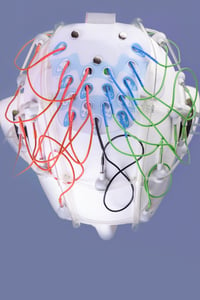 It is a recording of the electrical activity of the brain (brainwaves), using small sensors (electrodes) attached to the head. It detects very small electrical current that reflect neuronal activity of the brain. These signals are then amplified and can be graphed for visual inspection. The test time takes about an hour, results are available in about 24 hours, a trained technician is required to perform the test and a neurologist is required to read and interpret the results.
With 90% of EDs reporting overcrowding, wait times and lengths of stay a hot topic, why would this technology make sense in the ED?
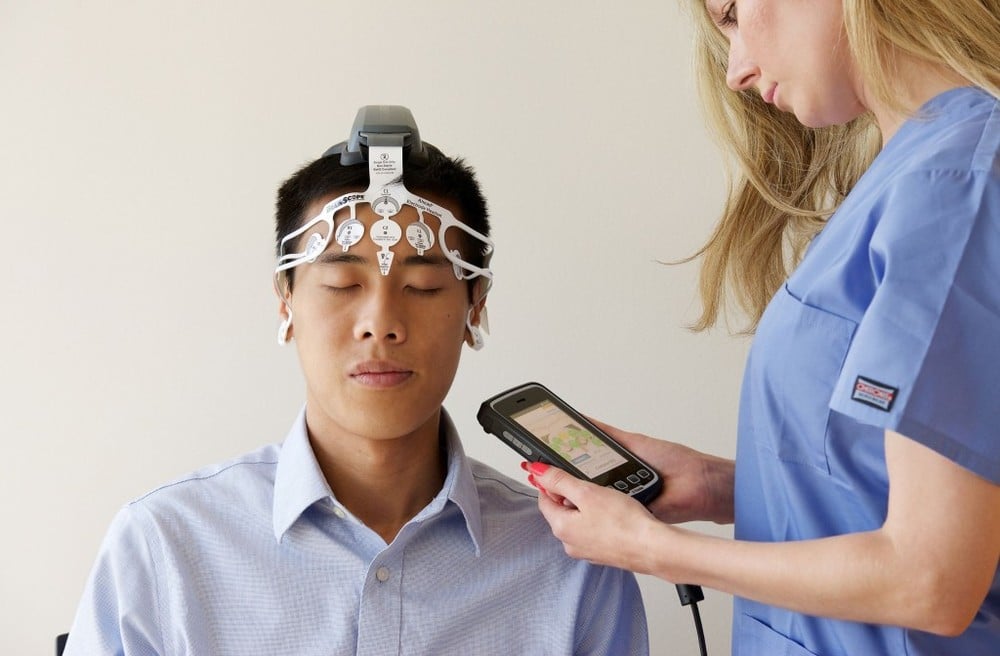
EEG has very high time resolution (milliseconds) and can capture physiological changes much better than other brain imaging tools (e.g., MRI or PET), making it uniquely suited to reflect the types of changes in brain activity that occur in mTBIs. The most vulnerable areas of brain injury in mTBIs include the frontal, temporal and frontotemporal regions. This allows for a limited set of electrodes compared to the traditional (pictured above) to be used in mTBI assessment.
BrainScope, through 12 years of research and development, has developed a proprietary set of algorithms, devices, and accessories that enables clinicians to rapidly assess mTBIs at the point of care with EEG. Using a single patient disposable headset attached to a handheld portable device, emergency department clinicians can have actionable results in 20 minutes or less.
.png?width=350&name=Brainscope%20stacked%201_headset%20(1).png) BrainScope’s technology uses quantitative electroencephalogram (qEEG) features. qEEG enables a large set of features which characterize the EEG signal to be identified which goes beyond those detected by visual inspection. Changes in the EEG can be described mathematically and deviations from the expected normal values (based on age) can be used to statistically describe abnormal signals in an individual and identify patterns of abnormalities distinctive of different disorders (e.g., mTBI)
BrainScope’s technology uses quantitative electroencephalogram (qEEG) features. qEEG enables a large set of features which characterize the EEG signal to be identified which goes beyond those detected by visual inspection. Changes in the EEG can be described mathematically and deviations from the expected normal values (based on age) can be used to statistically describe abnormal signals in an individual and identify patterns of abnormalities distinctive of different disorders (e.g., mTBI)
The proprietary algorithms provide multivariate EEG-based biomarkers to assess for structural and functional injury. The results are a composite of abnormal features found in the individual’s EEG and the likelihood that an individual with that pattern will be CT+ or CT-, or if they have brain function impairment such as that seen in concussion. This capability does not require a neurologist or electroencephalographer to “read” the brain waves.