Rapid, accurate, clinical decision support
Advanced digital signal processing of brain electrical activity data at the core of FDA cleared AI, machine learning derived algorithms, empower clinicians to rule out likelihood of intracranial hemorrhage & objectively assess for concussion

How BrainScope Works : Use of EEG based brain biomarkers to assess head injured patients and assist clinicians in their diagnosis
Exceptionally well validated
12 years of development funded in part by 8 Department of Defense studies and 2 GE/NFL Head Challenge grants
Sensitivity well above that of commonly used diagnostic tools for other medical conditions

BrainScope Structural Injury Classifier (SIC) was demonstrated to objectively identify the likelihood of an intracranial hemorrhage with 99% sensitivity to even the smallest amount of detectable blood (≥1 mL).
Here's what makes it work
Hardware
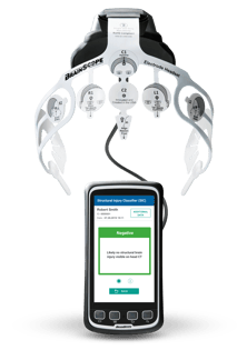
The handheld medical device acquires brain electrical activity data recorded from the proprietary 8-electrode disposable headset
AI Derived Biomarker Algorithms
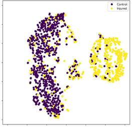
With brain electrical activity features as core inputs to machine learning classifier building methods, distinctive profiles of TBI are identified
A.I. Derived Biomarker Algorithms
Following an informed data reduction, selected EEG features are used as candidate inputs to A.I./machine learning based techniques (such as, genetic algorithms and LASSO Logistic Regression) for the derivation of the classifier algorithms. These sophisticated A.I. methodologies describe distinctive profiles or patterns of brain electrical activity used to determine the likelihood of structural brain injury (with sensitivity of 99% to the smallest amount of reliably detectable blood (>1 mL), and using the same EEG data, two separate algorithms for identifying the likelihood of concussion.
These algorithms have been demonstrated in independent FDA validation studies to have high accuracy. The algorithms provide a multivariate interpretation of the EEG data, which can be thought of as multivariate descriptors (unique profiles) which can be used to objectively identify the likelihood of a structural brain injury or (with separate algorithms) the likelihood of brain function impairment (concussion). This capability does not require a neurologist or electroencephalographer to read the brain waves and allows comparison to large populations of patients with closed head injury.
Read more in our Publications & Papers
BrainScope IP & patents
Virtual Patent Marking
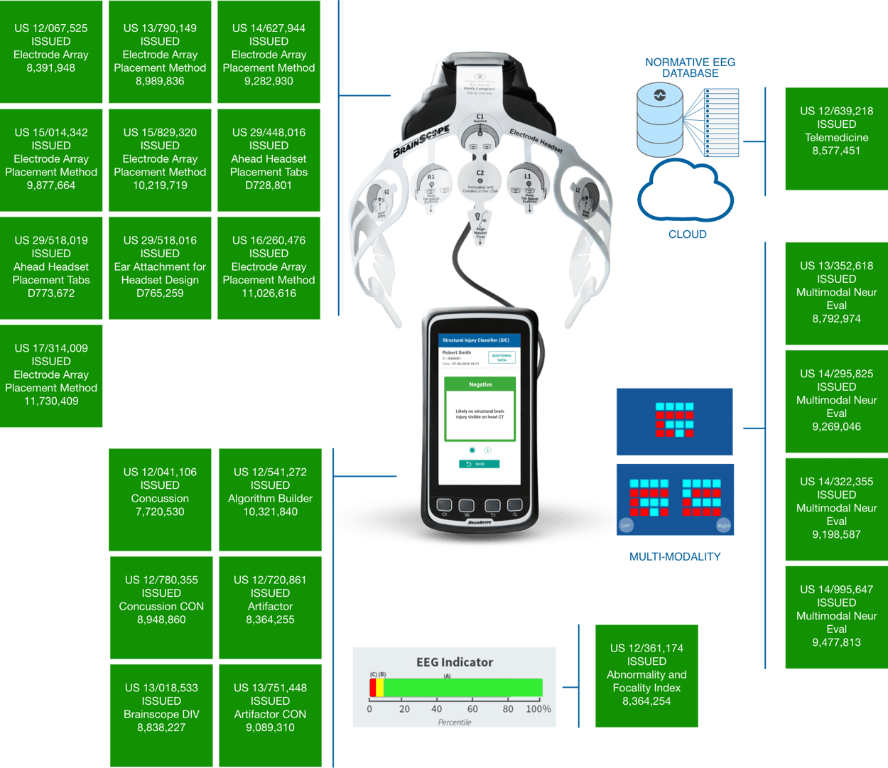
This document fulfills the U.S. patent marking requirement as defined under 35 U.S.C. §287. The products listed below are manufactured directly or indirectly by BrainScope Company, Inc. The U.S. patent or patents that provide patent protection of these products are listed in association with the product name. Additional U.S. and Non-U.S. patents may be pending.
BrainScope U.S. Patent Marking
Current assessment on the BrainScope device
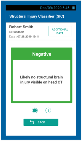
Structural Injury Classifier (SIC)
A multimodal AI derived algorithm that indicates the likelihood of being negative for brain bleed on a CT scan and identifies the need for further evaluation
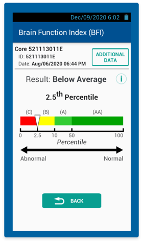
Brain Function Index (BFI) textpadding
A brain electrical activity based algorithm for the assessment of brain function impairment, obtained from the same recording used to compute the SIC—can aid in early clinical diagnosis of concussion and referrals
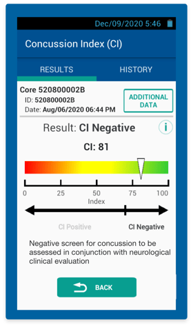
Concussion Index (CI)
An objective multimodal AI derived algorithm with brain electrical activity at its core—aids in clinical diagnosis of concussion
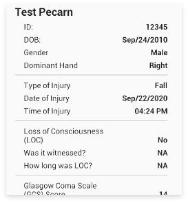
Digitized & Neurocognitive Clinical Assessments
Includes assessments commonly used by clinicians to assess head injured patients, including PECARN Decision Rule for pediatrics
Structural Injury Classifier
Indicated for use on patients 18-85 years, within 72 hours of injury, GCS 13-15
- Demonstrated to have 99% sensitivity to intracranial bleeds (>1 mL), a 98% negative predictive value (NPV), & specificity well above that of standard CT decision rules (CCHR & NOC)
- Potential to reduce head CT referrals by up to 60% when integrated into triage in the ED
BrainScope rapidly collects EEG data using a disposable electrode headset and then processes the data with specific clinical signs and symptoms using the BrainScope Structural Injury Classifier (SIC) algorithm. The algorithm extracts specific characteristics of the EEG signal from the artifact free (clean) data, uses age regression to remove the affect of age, and reports the result of the AI/machine learning derived algorithm, indicating the likelihood of presence or absence of structural brain injury.
In the FDA validation study, BrainScope’s SIC biomarker demonstrated extremely high sensitivity (99%) and negative predictive value (98%) for acute traumatic intracranial bleeds.
Read more in our Publications & Papers
Brain Function Index
Indicated for use on patients 18-85 years, within 72 hours of injury, GCS 13-15
- Scales significantly with the severity of clinical impairment
- Cannot be “gamed” or learned as can many of the standard, largely subjective, concussion assessment tools
Using the rapidly acquired EEG data, BrainScope also provides an objective assessment of brain function impairment, including concussion, with the Brain Function Index (BFI) algorithm. The BFI includes only EEG features, especially those that measure changes in “connectivity” between brain regions, reflecting the physiological changes seen in concussion.
The BFI is expressed as a percentile of a non head-injured population, from 0 to 100, with a lower score showing higher levels of impairment. This enables clinicians to make more confident clinical diagnoses of concussion using objective physiological data.
In the FDA validation study, the Brain Function Index was demonstrated to scale with severity of functional impairment: as the BFI goes down, the level of functional impairment increases.
Read more in our Publications & Papers
Concussion Index
Indicated for use on patients 13-25 years, GCS 15, within 72 hours
- Demonstrated in FDA validation study to have high accuracy for identifying likelihood of concussion
- Shown to be correlated with white matter integrity as seen with Diffusion Tensor Imaging
- Includes neurocognitive performance and vestibular information with EEG as highest contributor to the algorithm
The Concussion Index (CI) assessment incorporates rapidly acquired EEG data, cognitive performance testing, and specific clinical signs/symptoms into a multimodal algorithm to objectively assess concussion, with the largest contribution from EEG features only.
The CI is expressed as an index from 0 to 100 with a lower score indicating greater severity of injury. The CI assessment can aid in clinical decision making at the time of injury.
In the FDA Validation study the CI was demonstrated to have high accuracy in identifying the likelihood of concussion within 72 hours of injury. Injured patients with CIs less than or equal to threshold (70) are classified as Likely Concussed, and those with CIs greater than the threshold are classified as Not Likely Concussed.
Read more in our Publications & Papers
Neurocognitive & Digitized Assessments
Neurocognitive Assessments
The BrainScope® device includes a customizable battery of five cognitive performance tests, which are performed by the patient on the handheld device. These tests measure several cognitive functions including visuomotor reaction time, simple motor speed, working memory, and response control. Results can be calculated in comparison to normative data based on the non head-injured population of the same age and gender and in comparison to previous results for that patient using a reliable change index computation.
Digitized Assessments
BrainScope has digitized several standard clinical assessments commonly used by clinicians to assess head-injured patients. The digitized assessments are completed on the handheld and results for selected assessments are included in the patient PDF report and are displayed with their original intended formatting.
Digitized Assessments include:
• PECARN Decision Rule
• Sports Concussion Assessment Tool (SCAT5)
• Military Acute Concussion Evaluation (MACE 2)
• Near Point Convergence (NPC) and others.
Read more in our Publications & Papers
Our research team
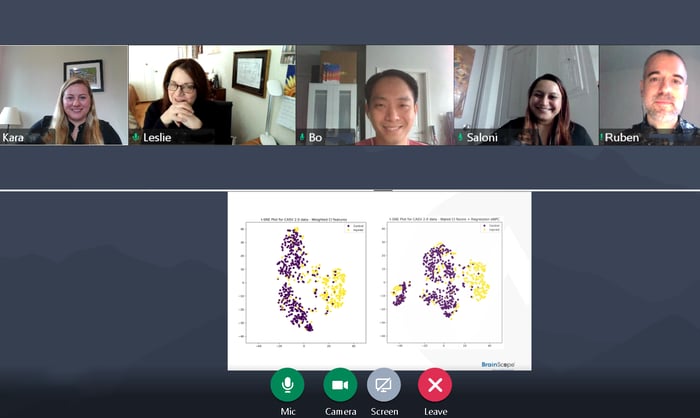
Led by our Chief Scientific Officer, Leslie S. Prichep, PhD, our vibrant research group has experience beyond traumatic brain injury into neurological conditions such as stroke, Alzheimer's disease, depression, and cognitive decline.
The BrainScope algorithms were developed by applying advanced AI/machine learning technology to extensive patient data, including EEG data, symptoms, CT scan and neurological test results. As the database grows, machine learning capabilities can identify additional data patterns, enhancing future technology, advancing the potential clinical application of head injury assessment, and identifying new indications for use, using the neurotechnology platform that has been developed by BrainScope over the past decade.
Dr. Prichep and her team continue to work on furthering our understanding of brain health.
Our research partners

University of Rochester Medical Center

Johns Hopkins Medicine

Texas Tech University

Washington University
Modal title
Cras mattis consectetur purus sit amet fermentum. Cras justo odio, dapibus ac facilisis in, egestas eget quam. Morbi leo risus, porta ac consectetur ac, vestibulum at eros.
Praesent commodo cursus magna, vel scelerisque nisl consectetur et. Vivamus sagittis lacus vel augue laoreet rutrum faucibus dolor auctor.
Our clinical collaborators
| Principal Investigator | Site |
|
Dr. John O’Neill |
Allegheny General Hospital |
|
Dr. Rosanne Naunheim |
Barnes Jewish Hospital/Washington University St. Louis |
|
Dr. John Garrett |
Baylor University |
|
Dr. Tamara Espinoza |
Emory University- Grady Hospital |
|
Dr. Joao Delgado |
Hartford Hospital |
|
Dr. Tracey Covassin |
Michigan State University |
|
Dr. Kenneth Podell |
Rice University |
|
Dr. Christopher Neville |
SUNY Upstate Medical University |
|
Dr. Robert Elbin |
University of Arkansas |
|
Dr. Douglas Casa |
University of Connecticut |
|
Dr. Neeraj Badjatia |
University of Maryland Medical Center/ R. Cowley Shock Trauma Hospital |
|
Dr. Gillian Hotz |
University of Miami |
|
Dr. Andrew Mayer |
University of New Mexico |
|
Dr. Jeffrey Bazarian |
University of Rochester Medical Center |
|
Dr. Susan Yeargin |
University of South Carolina |
|
Dr. Rebecca Lopez |
University of South Florida |
|
Dr. David Schnyer |
University of Texas at Austin |
|
Dr. J. Stephen Huff |
University of Virginia Medical Center Charlottesville, VA |
|
Dr. Elizabeth Jones |
UT-Houston Health Science Center Houston, TX |
|
Dr. Brian O’Neil |
Wayne State University/ Detroit Receiving Hospital & Sinai Grace Hospital |
Click here for a complete list of indications
EEG Brain Activity Database
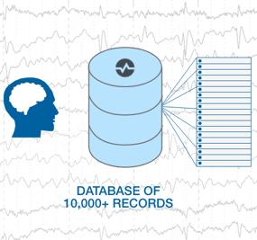
BrainScope’s database contains more than 14,000 evaluations from mTBI patients and normal subjects. 10,000 features that characterize the EEG signal are then extracted from each record and age-regressed relative to age expected normal values. These features are then used to power proprietary, A.I. derived FDA cleared algorithms to create objective biomarkers of brain injury.
BrainScope provides actionable results on the device in real time that also can be downloaded to a PDF. Real time streaming and recording of EEG data enable physicians to review raw EEG data and standard features, when desired.
Read more in our Publications & Papers
Hardware
EEG data is recorded from 8 electrode locations of the standardized expanded International 10-20 Electrode Placement System, referenced to linked ears. Data is acquired at a sampling rate of 1 kHz and all electrode impedances are below 10 kΩ. Amplifiers have a band pass filter from 0.3 to 250 Hz (3 dB points) and EEG data is down sampled to 100 Hz for feature extraction. Continuous monitoring of impedance occurs throughout the recording and is displayed on the acquisition screen so that the operator is notified, and acquisition is stopped if any lead rises above 10 kΩ. On-line artifact rejection algorithms are used to identify non-physiological signals and mark them for later removal prior to quantitative analyses.
Quantitative analysis of the artifact-free EEG (qEEG) data allows characterization of the signal into features used to describe brain activity, including measures of power, symmetry, coherence, phase, phase synchrony, complexity and others. Advances in signal processing have enriched these measures beyond those conventionally described, and approximately 10,000 features are derived from each EEG recording. The scientific literature demonstrates that the EEG of “normally functioning” individuals systematically changes over the life span and can be described by equations as a function of age. Deviations from the age expected normal values can be used to statistically describe abnormal signals in an individual, expressed as z-scores, removing the effect of age. Also, importantly, EEG has very high time resolution (milliseconds) and can capture physiological changes much better than other brain imaging tools (e.g., MRI or PET), making it uniquely suited to reflect the types of changes in brain activity that occur in mild traumatic brain injury (mTBI).
Read more in our Publications & Papers
